By Maria Jose Rodriguez | PhD and Associate of Apadrina la Ciencia
We hope you are all doing well, and you are enjoying the summer.
First of all, we would like to express our gratitude for your generous economic contributions.
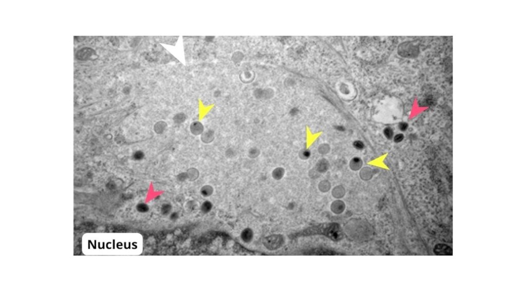
We also take this opportunity to share with you some information about one of the very useful instruments we use in research: Electron microscopes. They are undoubtedly one of the largest devices commonly used in science that a scientist may encounter during his or her career. And they serve, curiously, to reach to see the smallest components of life, the most unfathomable depths of everything that surrounds us.
The phrase "Seeing is believing" is a good description of the powerful impact that electron microscopy has had on our understanding of matter, as demonstrated by the number of Nobel Prizes awarded for work that focused on the use of this technique.
Electron microscopy, which has been instrumental in the development of cell and molecular biology over the past decades, is living a new golden age thanks to the advances that have been made in recent years. While the importance of the construction of the electron microscope in 1931 by Ernst Ruska and Max Knoll was highlighted by the award to Ruska of the Nobel Prize in Physics in 1986, improved sample preparation techniques, instrumental development and computational improvements have brought electron microscopy to the forefront of structural biology research. And this has resulted in a new Nobel Prize being awarded: the Nobel Prize in Chemistry in 2017 to the driving forces behind electron cryomicroscopy, Jacques Dubochet, Joachim Frank and Richard Henderson.
The increase in the number of applications of the transmission electron microscope for molecular and cellular studies has grown dramatically from the 1940s to the present day (Harris, 2015). In fact it is a fundamental tool in the study of biological systems, from proteins to whole cells or tissues, providing most of the existing knowledge about the ultrastructural organization of tissues, cells and subcellular organelles.
In addition to its usefulness in basic research, electron microscopy has had an enormous impact on diagnostic microbiology since the late 1960's. Viruses from different families have different morphology and this variation is the basis for virus identification by electron microscopy. The 1970s and 1980s were a boom period for electron microscopy, which led to the discovery of a large number of new clinically important viruses such as adenoviruses, enteroviruses, paramyxoviruses, etc. There are currently 103 recognized virus families, of which 22 infect humans (Goldsmith, 2014) and it is postulated that there are millions of viruses occupying all terrestrial and marine ecosystems capable of infecting all types of living beings: animals, plants, fungi and bacteria.
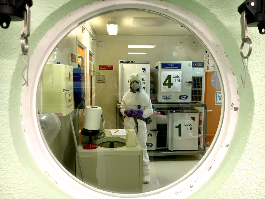
As we are unfortunately experiencing nowadays, the emergence of new viruses with pandemic potential or the use of viruses and other microorganisms as a threat of bioterrorism is not something that is confined to the realm of storytelling. The development of molecular techniques such as RT-PCR or ELISA has replaced electron microscopy in many areas of virus diagnosis in recent years. However, electron microscopy continues to offer enormous advantages in several key areas: speed of diagnosis, critical for initiating appropriate treatment and/or containment protocols without waiting for additional testing; the potential to detect, with a single test, any viral pathogen and even multiple pathogens present in a single sample; initial identification in the case of an unknown pathogen (molecular tests cannot be applied without prior information on the infectious agent); utility in the investigation and diagnosis of emerging viruses. In addition, electron microscopy remains a vital part of national emergency response programs in many countries and is a first-line diagnostic service for any bioterrorism threat (Goldsmith and Miller, 2009).
Please help us spread the word further, among family, friends and colleagues.
By donating your money to Apadrina la Ciencia, you are contributing to projects that advance the knowledge about our environment and also supporting the development of early career researchers in Spain.
Thank you all again for your support!
- Goldsmith, C.S.; Miller, S.E. (2009). Clinical Microbiology Reviews. 22(4): 552-563
- Harris, J.R. (2015). Archives of Biochemistry and Biophysics. 581: 3-18
Project reports on GlobalGiving are posted directly to globalgiving.org by Project Leaders as they are completed, generally every 3-4 months. To protect the integrity of these documents, GlobalGiving does not alter them; therefore you may find some language or formatting issues.
If you donate to this project or have donated to this project, you can receive an email when this project posts a report. You can also subscribe for reports without donating.
Support this important cause by creating a personalized fundraising page.
Start a Fundraiser